Aortic coarctation
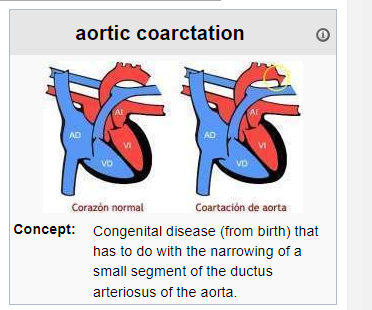
Aortic coarctation or coarctation of the aorta . It is a congenital disease (from birth) that has to do with the narrowing of a small segment of the ductus arteriosus of the aorta. The aorta is the large artery that carries oxygen-rich (red) blood from the left ventricle to the body.
Summary
[ disguise ]
- 1 Course of the aorta vein
- 2 There are three types of aortic coarctation
- 3 Symptoms and signs
- 4 Causes
- 5 Tests and exams
- 6 Treatment
- 7 Forecast
- 8 Possible complications
- 9 Sources
Course of the aorta vein
It has the shape of a staff; The first section rises toward the head (ascending aorta), then curves into a “C” shape as smaller arteries connected to it carry blood to the head and arms (aortic arch). After the bend, the aorta becomes straight again and moves downward toward the abdomen , carrying blood to the lower part of the body (descending aorta).
There are three types of aortic coarctation
- Preductal coarctation: The narrowing is proximal to the ductus arteriosus. Blood flow to the aorta that is distal to the narrowing is dependent on the ductus arteriosus; therefore, very severe coarctations can be life-threatening. Preductal coarctation results when an intracardial anomaly during fetal life decreases blood flow through the left chamber of the heart, producing hypoplasia in the development of the aorta. It is observed in approximately 5% of patients with Turner Syndrome.
- Ductal coarctation: Narrowing occurs at the insertion of the ductus arteriosus. This type usually appears when the ductus arteriosus closes (at birth).
- Postductal coarctation: The narrowing is distal to the insertion of the ductus arteriosus. Even with an open ductus arteriosus, blood flow to the lower part of the body is irregular. This type is the most common in adults. It is associated with the finding of notches on the chest x-ray at the level of the stenosis and also at the level of the rib cage (due to increased pressure at the level of the intercostal arteries), hypertension in the upper extremities and weakness in the pulses in the lower extremities. Postductal coarctation is probably the result of the extension of a muscular artery (the ductus arteriosus) into the elastic artery (aorta) during fetal life, when contraction and fibrosis close the ductus arteriosus during birth. produces narrowing of the aortic lumen.
Symptoms and signs
Symptoms depend on the amount of blood that can flow through the artery. About half of newborns with this problem will have symptoms in the first days of life. In milder cases, symptoms may not appear until the child has reached adolescence. Symptoms include:
- Dizziness or fainting
- Difficulty breathing
- Throbbing headache
- Chest pain
- Cold hands and feet
- Nosebleed
- Leg cramps with exercise
- High blood pressure ( hypertension) with exercise
- Decreased exercise capacity
- Development delay
- Poor growth
Note: There may be no symptoms. This narrowing causes an unmistakable heart murmur, irregular high blood pressure in the arms, and reduced blood flow in the abdomen, pelvis, and legs. It increases the stress on the left ventricle, which usually results in its enlargement.
Causes
The aorta carries blood from the heart to blood vessels that supply the body with blood and nutrients. If part of this artery narrows, it is difficult for blood to pass through it.
Aortic coarctation is more common in people with certain genetic disorders, such as Turner syndrome. However, this condition can also be caused by congenital defects of the aortic valves.
Aortic coarctation is one of the most common heart conditions that are present at birth (congenital heart conditions). It is usually diagnosed in children or adults under 40 years of age.
Coarctation of the aorta can be seen with other congenital heart defects, such as:
- Bicuspid aortic valve
- Defects in which only one ventricle is present
- Ventricular septal defect
Tests and exams
The doctor will perform a physical exam and take blood pressure in your arms and legs.
- The pulse in the femoral (groin) area or feet will be weaker than the carotid (neck) pulse and sometimes may not be felt at all.
- Blood pressure in the legs is generally weaker than in the arms. Blood pressureis usually higher in the arms after breastfeeding.
- The doctor listens to the heart and will check for the presence of murmurs. People with aortic coarctation have a high-pitched murmur that can be heard from the back. Other types of murmurs may also occur.
- Echocardiography
- chest x-ray
- CT scan of the heart
- MRIor magnetic resonance angiography
- Cardiac catheterization
Both Doppler ultrasound and cardiac catheterization can be used to see if there are any differences in blood pressure in different areas of the aorta.
Treatment
Treatment can be conservative if the patient is asymptomatic, but the use of surgery is the norm if aortic narrowing causes hypertension. The first operation was performed by Clarence Crafoord in Sweden in 1944. In some cases, angioplasty can be used to dilate the artery. Surgical reconstruction, or replacement of the constricted area, is more effective in younger people than in older patients.
Forecast
Coarctation of the aorta can be cured with surgery and symptoms improve quickly after surgery. However, there is an increased risk of death due to heart problems among people who have had aortic repair. Without treatment, most people with this condition die before age 40. For this reason, doctors generally recommend that the patient have surgery before the age of 10. In fact, most of the time, surgery to repair aortic coarctation is performed during the first year of life. Narrowing or coarctation of the artery can return after surgery and is more likely in people who had surgery when they were newborns.
Possible complications
Before, during and shortly after surgery include:
- aortic aneurysm
- Aortic dissection
- Aortic rupture
- intracerebral hemorrhage
- Endocarditis(heart infection)
- Heart failure
- Hoarseness caused by injury to the laryngeal nerve
- Impaired renal function
- Paralysis of the lower half of the body (rare complication of surgery to repair coarctation)
- Premature development of coronary artery disease (CAD)
- Severe arterial hypertension
- stroke
Long term they include:
- Continuous narrowing of the aorta
- Endocarditis (heart infection)
- Arterial hypertension